
A guide to medical imaging
X-ray, Ultrasound, CT, MRI and PET-CT: What’s the Difference?
Understand what each scan can show, how it works and why the right choice depends on the clinical question—not simply the most detailed image.
X-ray, Ultrasound, CT, MRI and PET-CT: Which Scan Does What?
Estimated reading time: 8 minutes.
Understanding the differences between X-ray, ultrasound, CT scan and MRI can make conversations about imaging less confusing. PET-CT adds another perspective by showing biological activity alongside anatomy. Whether you’re investigating a possible fracture, a soft-tissue concern or persistent back pain, the most useful test depends on the question a healthcare professional needs to answer. Sometimes an examination or another test is more appropriate than a scan. This guide explains the strengths, limitations and radiation considerations of each method without suggesting that one scan is right for every person.
Five scans, five different clinical roles
These imaging methods answer different questions. Your symptoms, examination findings and medical history help a clinician decide whether imaging is needed and which type may be appropriate.
X-ray: A first look at bones
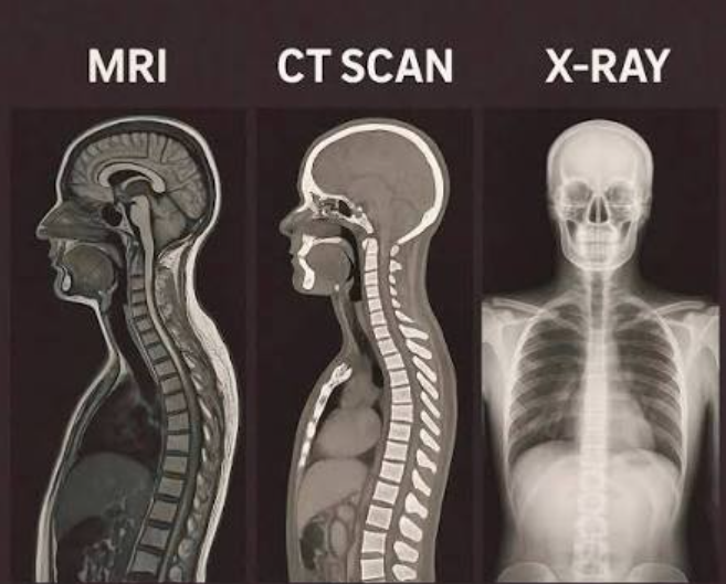
X-rays pass ionising radiation through the body to create flat images. They’re commonly used to assess suspected fractures and joint changes, but show muscles and ligaments less clearly than some other methods.
Ultrasound: Soft tissues in motion
Ultrasound uses high-frequency sound waves to produce live images without ionising radiation. It can assess tendons, muscles and some joints, although image quality depends on the area examined and the operator’s skill.
CT: Fast cross-sectional detail
A CT scan uses X-rays and computer processing to build detailed cross-sectional images. It is often useful in trauma, complex fractures and assessments of internal organs when answers are needed quickly. CT uses ionising radiation; some examinations also use contrast material.
MRI and PET-CT: Different kinds of detail
MRI uses a strong magnetic field and radio waves—not ionising radiation—to show the brain, spine and soft tissues such as discs, ligaments and muscles. PET-CT combines a radioactive tracer that reveals biological activity with CT images showing anatomy; it is used particularly in certain cancer investigations. PET-CT involves ionising radiation and is not a routine test for muscle or joint injuries.
CT scans: fast, detailed views inside the body
A CT scan uses X-rays taken from multiple angles to create detailed cross-sectional images of bones, blood vessels and internal organs. Its speed makes it particularly useful when clinicians need to assess serious trauma, complex fractures or possible internal injuries urgently. CT uses ionising radiation, so its benefits must be weighed against radiation exposure. Some examinations also use a contrast material to make particular tissues or blood vessels easier to see; your healthcare team can explain whether contrast is appropriate for you.
MRI: a closer look at soft tissues
MRI uses a strong magnetic field and radio waves—not ionising radiation—to produce detailed images of the brain, spinal cord, intervertebral discs, ligaments, cartilage and muscles. For someone exploring assisted stretching because of persistent discomfort or restricted movement, MRI may help answer a specific clinical question when a qualified healthcare professional considers it necessary; it is not automatically needed for every muscle ache or backache. Scans generally take longer than CT, may cost more and may be less readily available. The enclosed scanner can feel uncomfortable if you experience claustrophobia, and certain implants or metal devices require careful safety screening before an examination. MRI findings must be considered alongside symptoms, medical history and a physical assessment.
PET-CT: biological activity meets anatomical detail
A PET-CT scan combines two kinds of information: PET uses a small amount of radioactive tracer to show metabolic or other biological activity, while CT provides detailed anatomical images that help locate those findings. Clinicians may use PET-CT for certain cancer investigations, including diagnosis, staging and monitoring response to treatment, as well as selected other medical conditions. It is not a routine scan for everyday muscle or joint concerns encountered in assisted stretching. Because the tracer is radioactive and the CT component uses X-rays, radiation exposure is an important consideration when a healthcare professional decides whether the test is warranted.
Medical scans at a glance
X-ray and CT
Both use ionising X-rays. X-ray gives quick, flat images often used for suspected fractures; CT builds detailed cross-sections for complex injuries and internal organs. CT generally involves more radiation, and some scans require contrast.
Ultrasound and MRI
Neither uses ionising radiation. Ultrasound uses sound waves to examine moving tissues, including some muscle and tendon injuries; results can depend on the operator. MRI uses magnetic fields and radio waves for detailed soft-tissue images but takes longer and requires implant safety checks.
PET-CT
A radioactive tracer highlights biological activity while CT locates findings anatomically. It is used in selected cancer assessments and other conditions, not routinely for muscle or joint injuries. Both the tracer and CT contribute to ionising-radiation exposure.
Which scan might be considered?
Suspected fracture
An X-ray is often the first investigation when a fracture is suspected, although examination and the circumstances of the injury guide the decision.
Possible muscle tear
Ultrasound may help assess some muscle tears, especially when real-time imaging would answer a specific clinical question.
Suspected tendon injury
Ultrasound or MRI may be considered, depending on the tendon, the suspected injury and whether imaging would change care.
Complex trauma
CT can provide detailed images quickly when clinicians need to assess complex fractures or injuries after significant trauma.
Possible internal injury
CT may be appropriate for certain suspected internal injuries, sometimes with contrast material to make structures easier to assess.
Persistent spinal symptoms
MRI may be considered when ongoing back or neck symptoms suggest a spinal condition and the result could inform treatment.
Suspected nerve-related condition
When symptoms and examination indicate possible nerve involvement, a clinician may consider MRI; it is not needed for every backache.
Selected cancer investigations
PET-CT may help with staging or treatment monitoring in certain cancers. These are examples, not fixed rules: other tests or no imaging may be more appropriate.
Does a more advanced scan mean a better diagnosis?
Not necessarily. The best test is the one that helps answer a specific clinical question. An X-ray may be the right starting point for a suspected fracture, while MRI might add detail only when it would affect what happens next. More detailed imaging can take longer, cost more or introduce considerations such as radiation exposure, contrast material or implant safety. For muscle or joint concerns raised during assisted stretching, a qualified healthcare professional can advise whether imaging is useful at all.


Why do symptoms and examination matter?
A scan shows selected structures, not the full experience of pain or movement difficulty. Clinicians consider the images alongside your symptoms, medical history and physical examination to judge whether a finding is relevant.
Can a scan show something unrelated to my pain?
Yes. Some changes appear on imaging in people who have no symptoms. A reported abnormality does not automatically identify the cause of discomfort, so its location and clinical context matter.
When might imaging not be needed?
If a scan is unlikely to change the next step, a clinician may recommend monitoring or other care first. Discuss persistent, worsening or concerning symptoms with a qualified healthcare professional rather than choosing a scan based on how advanced it sounds.
Frequently Asked Questions About Medical Scans
These answers explain common imaging decisions, but your symptoms and clinical assessment determine what is appropriate for you.
Is MRI better than CT?
Neither is always better. MRI is often useful for detailed soft-tissue images without ionising radiation; CT can provide detailed images quickly, especially in emergencies. The best choice depends on the clinical question.
Can an ultrasound detect a muscle tear?
Ultrasound can identify some muscle tears and assess tissue while it moves, but results depend on the area examined and the operator’s skill. A clinician may recommend another approach if ultrasound cannot answer the question clearly.
Can an X-ray show muscle or ligament damage?
X-rays primarily show bones. They generally do not show muscles or ligaments in detail, although they can reveal a fracture or other bone change associated with an injury.
Does an MRI use radiation?
MRI uses a strong magnetic field and radio waves, not ionising radiation. Tell your healthcare team about implants or metal in your body before a scan so they can check MRI safety.
Why might a doctor not order a scan immediately?
A medical history and physical examination may provide enough information to guide initial care. A scan is most helpful when its result could change a diagnosis or treatment plan; some findings can appear on scans without causing symptoms.
Do I need an MRI for neck or back pain?
Not necessarily. Many cases of neck or back pain do not require an immediate MRI. A clinician may consider imaging if symptoms, examination findings or warning signs suggest it could help guide care.
Explore Imaging Guidance and Related Reading
On The Rel Therapy Website
Medical Imaging References
Questions to Discuss With a Clinician
The Right Scan Depends on the Question
X-ray, ultrasound, CT, MRI and PET-CT each serve different purposes. The most useful scan is the one that helps answer the relevant clinical question—not necessarily the most advanced or expensive. Discuss your symptoms, concerns and imaging options with a qualified healthcare professional. This article is for general educational purposes only and does not replace professional medical advice. If you have symptoms or concerns about a medical condition, consult a qualified healthcare professional.